


Predict Intestinal Permeability from Direct Membrane Interaction
The TRANSIL Intestinal Absorption Kit directly measures drug affinity to phospholipid membranes and predicts intestinal permeability (Pint) and tissue distribution from mechanistic membrane partitioning behavior.
Not all lipophilicity is biologically relevant. LogP and logD measure solvent partitioning.
The TRANSIL assay measures partitioning into biological membranes. By capturing the molecular interaction between drugs and phospholipid bilayers, membrane affinity becomes a mechanistically meaningful predictor of intestinal permeability and tissue distribution.
Key benefits:
- Mechanistic measurement of membrane affinity
- Prediction of intestinal permeability (Pint)
- Insight into tissue distribution (VD)
- Rapid equilibrium assay (minutes instead of hours)
Lipophilicity is a Poor Proxy for Membrane Interaction
Lipophilicity metrics such as logP or logD are widely used to estimate permeability, but they measure partitioning between water and artificial organic solvents like octanol. Biological membranes are fundamentally different. They consist of structured phospholipid bilayers with charged headgroups, hydrophobic cores, and dynamic molecular organization. As a result, compounds with similar logP values often interact very differently with real membranes. Since passive intestinal absorption is governed by partitioning into phospholipid membranes, solvent-based lipophilicity scales provide only an indirect and often misleading proxy.
PAMPA assays attempt to address this limitation by introducing a lipid barrier. However, PAMPA membranes consist of a liquid lipid layer rather than a structured phospholipid bilayer. Lipids are dissolved in organic solvents and immobilized in filter supports, forming a fluid lipid film that lacks the ordered architecture of biological membranes. Consequently, PAMPA membranes cannot fully reproduce the molecular interactions that govern drug partitioning into biological membranes.
The TRANSIL Intestinal Absorption Kit overcomes these limitations by using packed phosphatidylcholine bilayers with natural membrane fluidity. This creates a membrane environment that closely resembles biological membranes and allows direct measurement of compound partitioning into phospholipid bilayers — the fundamental interaction driving passive intestinal permeability and tissue distribution.
The Biological Basis of Intestinal Absorption and Tissue Distribution
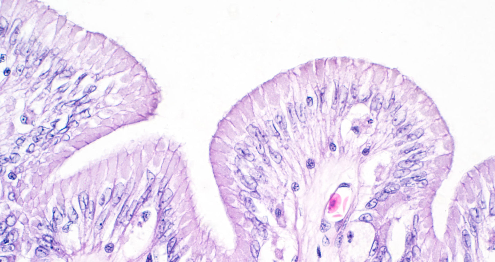
Absorption of orally administered drugs across the intestinal epithelium is primarily governed by passive diffusion through phospholipid membranes. To enter systemic circulation, compounds must first partition from the aqueous intestinal lumen into the lipid bilayer of epithelial cell membranes and subsequently diffuse across this membrane barrier. The efficiency of this process is therefore largely determined by the compound’s affinity for phospholipid membranes, which reflects the molecular interactions between the drug and the lipid bilayer. As a result, membrane partitioning is a key determinant of intestinal permeability and also contributes to tissue distribution once compounds enter systemic circulation.
The TRANSIL Intestinal Absorption Kit
The TRANSIL Intestinal Absorption Kit directly measures the affinity of compounds for phosphatidylcholine membranes, the dominant lipid class in intestinal epithelial cell membranes. In the assay, test compounds are incubated with increasing amounts of immobilized phospholipid bilayers, allowing rapid equilibrium partitioning between the aqueous phase and the membrane phase. From the resulting distribution of the compound, the membrane affinity coefficient is determined. This experimentally measured membrane affinity captures the molecular interaction between drugs and biological lipid bilayers and can be used to predict intestinal permeability (Pint) as well as contribute to the estimation of volume of distribution. By measuring the fundamental process underlying passive membrane permeation, the TRANSIL approach provides a mechanistically relevant alternative to solvent-based lipophilicity measurements.
How the Assay Works
The assay quantifies membrane affinity by measuring how test compounds distribute between the aqueous phase and immobilized phosphatidylcholine membranes. After incubation, membrane beads are separated and the remaining compound concentration in the supernatant is determined. The change in concentration across wells containing different membrane amounts is then used to calculate the membrane affinity coefficient.
The assay determines membrane affinity through the following equilibrium partitioning workflow:
- Compound is added to wells containing increasing amounts of membrane- coated beads
- Compound partitions between membrane and aqueous phase
- Supernatant concentration is quantified
- Membrane affinity is calculated from the equilibrium distribution
- Intestinal permeability (Pint) and volume of distribution (VD) are predicted from the measured membrane affinity

Features and Benefits
- Direct measurement of membrane affinity to phosphatidylcholine bilayers — Provides a mechanistically relevant descriptor of intestinal permeability and tissue distribution.
- Biologically realistic phospholipid membranes — Captures true drug–membrane interactions that are not represented by solvent-based lipophilicity measurements.
- Prediction of intestinal permeability (Pint) — Enables early identification and optimization of compounds with favorable oral absorption.
- Contribution to prediction of volume of distribution (VD) — Provides insight into tissue partitioning and pharmacokinetic behavior during lead optimization.
- Integrated data quality assessment (TRANSIL Quality Index, TQI) — Multiple internal quality control parameters automatically evaluate data reliability and help identify experimental artifacts.
- Rapid equilibrium partitioning assay — Generates results within minutes rather than hours required for many permeability assays.
- Cell-free assay format — Eliminates variability caused by transporters, metabolism, and cell culture conditions.
- Simple centrifugation-based separation — Allows straightforward workflows without specialized permeability equipment.
- 96-well plate format — Enables high-throughput screening of discovery compounds.
- Quantitative mechanistic parameter (membrane affinity coefficient) — Provides a transferable physicochemical descriptor that can be used in pharmacokinetic modeling.
Validation Against Human Absorption Data
A study comprising 126 chemically diverse marketed drugs demonstrated that membrane affinity is a reliable predictor of passive intestinal absorption. Predicted fractions absorbed derived from membrane affinity measurements show good agreement with reported human absorption values across most compounds, indicating that passive membrane partitioning is the dominant determinant of oral drug absorption (Figure 1). Compounds that deviate strongly from the prediction are typically influenced by active transport processes, highlighting the contribution of influx or efflux transporters beyond passive permeability.

-
Is membrane affinity more meaningful than measuring lipophilicity as LogP or LogD?
-
How do we know that the immobilized membranes retain their natural fluidity?
An important goal for designing the TRANSIL kits was to model a compound’s interaction with membranes as close as possible. That requires that the membranes retain their natural fluidity. To achieve that we immobilize single membrane bilayers on porous silica beads such that the membranes float on a thin water layer. The immobilization conditions have been optimized such that both differential scanning calorimetry (c.f. figure 3) and NMR spectrometry (c.f. figure 4) show very similar fluidity patterns.

Figure 3: Differential scanning calorimetry comparing free floating liposomes (phosphatidylcholine vesicles) with immobilized TRANSIL beads. Both vesicles and beads melt at the same temperature, which indicates that they have the same structure and their fluidity is comparable.
Figure 4: Comparison of H2-NMR spectrums of free floating membrane vesicles (blue) and TRANSIL bead supported membranes (red). Energy peaks of rotation and flipping of phospholipids occurs at the same frequencies (kHz) in both supported and unsupported membranes. This indicates that the TRANSIL membrane support beads stabilized the phosphatidylcholine membranes such that they retain their natural fluidity. -
How does the TRANSIL Intestinal Absorption Kit work?
The TRANSIL Intestinal Absorption Kit measures the affinity of a test item to immobilized phosphatidylcholine membranes with natural membrane fluidity. This membrane affinity is a partitioning coefficient of drug between membrane and buffer. It is defined as the concentration of drug in membrane (cl) over the concentration of drug in buffer (cb):

The membrane affinity is calculated from the assay data using the mass balance equation:

which is rearranged such that the membrane affinity can be determined from the slope of plotting the ratio of total amount of drug (nt) over remaining concentration in supernatant (cb) against the lipid membrane volume present in each well:

-
What are the main quality control measures applied in TRANSIL assays?
-
How long does it take to run the assay?
-
How many compounds can be analyzed with one plate?
-
Are TRANSIL assay kits supplied in low-binding plates?